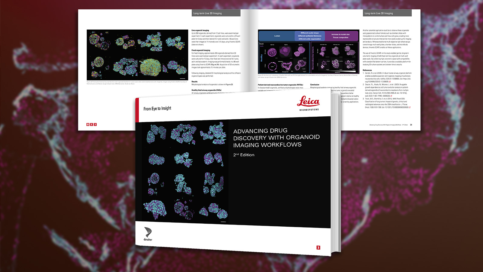
Complex In Vitro Models (CIVMs) such as organoids are becoming increasingly important in early drug discovery and translational research, supporting the shift toward more predictive, human-relevant data in line with evolving regulatory expectations.
However, extracting meaningful information from these models is not straightforward. Their size, heterogeneity and scalability, combined with sensitivity to handling and photodamage—can make robust imaging and characterization challenging.
This eBook provides a practical, workflow-oriented guide to organoid imaging and analysis—helping you navigate key challenges and compare approaches to select the right fit for your experiments. It is supported by real-world examples from biotech and translational research.
What you’ll learn:
- Approaches for tracking cell development, behaviour, and interactions in live organoids using gentle long-term imaging strategies
- Considerations for scaling experiments with multi-well plate-compatible workflows
- Strategies for imaging cleared samples to visualize structures deep within large organoids
- Options for 3D imaging of thick and dynamic samples with high-speed and high-resolution
Case studies include:
- Characterizing healthy vs. tumor airway organoids at scale
- Imaging extended sections of uterine organoids at high-resolution and high-speed
- Tracking cell lineage and development of intestinal organoids over time
Find an approach that fits your application
Gain a clearer understanding of how different imaging approaches can be combined to answer specific experimental questions, and where each can add unique value within organoid imaging workflows.