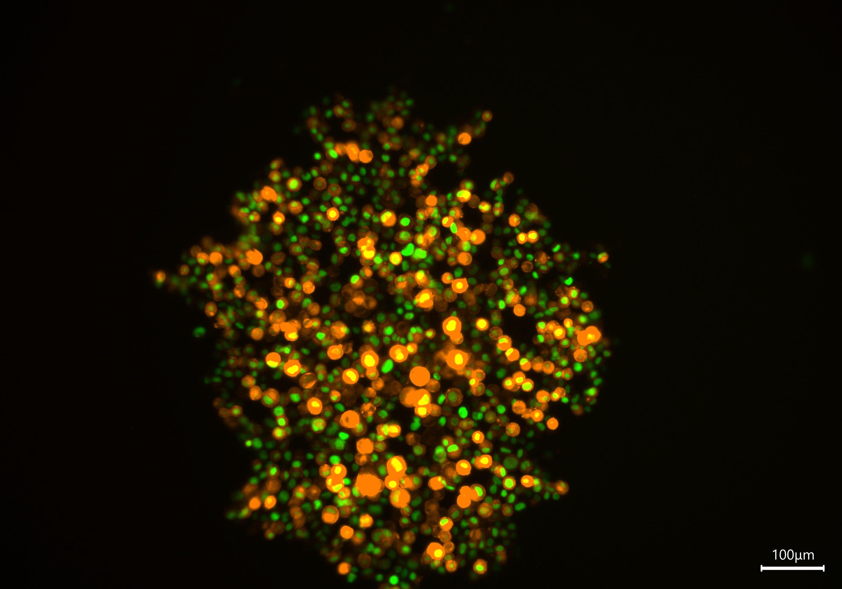
T47D breast cancer cell spheroid - Formation (Day 0). Cells were labeled for nuclei (Histone2B-eGFP) and F-actin (LifeActmCherry); scale bar 100 μm. Image acquired using Mateo FL.
Monitoring organoid growth can be challenging, as removing samples from their culture systems often risks compromising their structural integrity. This application note highlights non‑invasive, efficient digital microscopy approaches for real‑time observation directly within culture systems, minimizing disturbance while delivering high‑quality imaging.
____________________________________________________________________________________________
Imaging Complex Human-Relevant Models for Early Drug Discovery - Technology Showcase
25th June 2026
Join Leica Microsystems and leading experts as they explore how organoids are being used across early drug discovery and translational research workflows, the challenges associated with working with increasingly complex models, and emerging imaging approaches designed to address some of these challenges.