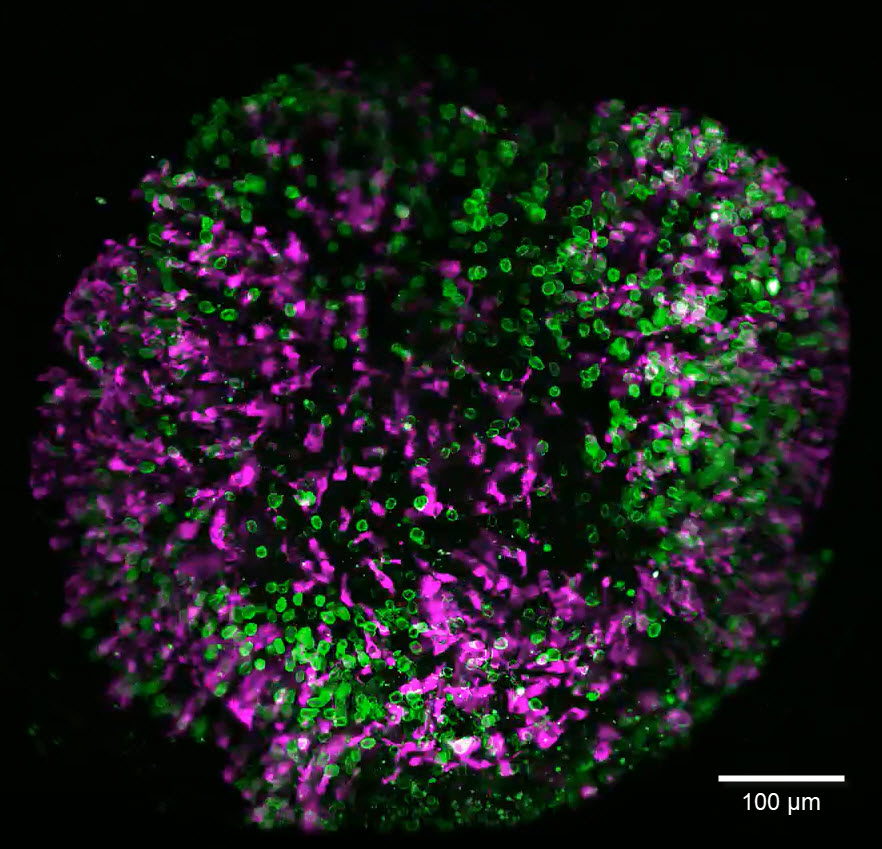
Brain organoids labeled with lamin (green) and tubulin (magenta), acquired using Viventis Deep. Courtesy of Akanksha Jain, Treutlein Lab ETH-DBSSE Basel (Switzerland)
Develop deeper, more translatable, insights into organoid and spheroid models used in drug discovery and disease research
Learn how advanced microscopy techniques and integrated workflows can help maximize the potential of physiologically relevant 3D model systems — spanning routine cell culture monitoring and pre‑screening through to high‑resolution, long‑term live cell imaging and AI‑based image analysis.
These approaches can contribute to the advancement of new approach methodologies (NAMs).
Why download the eBook?
- Discover strategies to address common imaging challenges associated with dense, multicellular 3D systems
- Explore real‑world case studies where microscopy is used to investigate drug uptake, disease models, single‑cell fate tracking in organoids and spheroids, and more
- See how advanced imaging techniques provide complementary perspectives on lung, intestinal, endometrial, and breast cancer organoids
____________________________________________________________________________________________
Imaging Complex Human-Relevant Models for Early Drug Discovery - Technology Showcase
25th June 2026
Join Leica Microsystems and leading experts as they explore how organoids are being used across early drug discovery and translational research workflows, the challenges associated with working with increasingly complex models, and emerging imaging approaches designed to address some of these challenges.