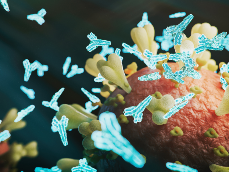
Understanding how and where an antibody binds to its target antigen is central to developing effective biologics. Yet, conventional techniques such as, alanine scanning, or HDX-MS, only provide partial or indirect insights into these critical epitope–paratope interactions.
This is where ATEM redefines the landscape. By applying Cryo-Electron Microscopy (Cryo-EM)–based High-Throughput Epitope Mapping, ATEM enables direct 3D visualization of antigen–antibody complexes. This breakthrough approach empowers pharmaceutical scientists to optimize binding, improve selectivity, and accelerate candidate development with unprecedented structural clarity.
Through a fully integrated workflow spanning sample preparation, imaging, and AI-powered computational analysis, ATEM delivers 3D structural data that expose the true structure–function relationships driving therapeutic efficacy.
Our High-Throughput Epitope Mapping Service transforms antibody characterization by providing directly interpretable and unambiguous 3D data that reveal exactly how antibodies (Fabs, scFvs, or mAbs) engage their targets. Leveraging advanced cryo-EM instrumentation and proprietary image analysis, we define contact residues, binding orientations, and conformational epitopes that underpin biological activity.
At ATEM we provide our collaborators with a clearly milestoned workflow and work-packages. This keeps you in control of the process and the budget. ATEMs 3D epitope mapping screening-package for example enables you to map up to five structures in just three weeks. This reduces ambiguity, cost, and timelines compared to traditional workflows.
Key Benefits:
· Direct Visualization: Resolve Fab–antigen interfaces in 3D, no inference required.
· Applicable to Complex Targets: Including large, flexible, or membrane proteins.
· Quantitative Data Outputs: Define footprint areas, contact residues, and structural overlaps.
· Fast Turnaround: Integrated sample-to-structure 3 weeks workflow reduces development cycles.
· Expert Oversight: Every project guided by experienced structural biologists.
Applications:
· Validate lead antibodies early in discovery.
· Identify overlapping or non-competitive epitopes for combination therapies.
· Support affinity, maturation, and rational design.
· Strengthen IP and regulatory filings with structural data.
· Visualize antigen conformational flexibility or allosteric sites.
Insights Report Introduction: Bridging the Resolution Gap
Traditional epitope mapping methods like HDX-MS and alanine scanning are valuable for early triage but fall short for lead selection due to limited resolution and ambiguous outputs. On the other end, X-ray crystallography delivers atomic detail but is slow, costly, and often incompatible with complex or glycosylated targets.
ATEM’s EpiCore™ computational platform closes this “resolution gap” by offering high-resolution insights without the need for crystallization, capturing antibody–antigen complexes in their native state.
Report Outcomes:
· Native-state visualization of complexes, preserving conformational changes and post-translational modifications.
· High-confidence epitope definition (4–10 Å for rapid screening; < 4 Å for residue-level detail).
· Seamless integration with AI-driven lead prediction and structure-based design workflows.
· Validated cost-efficiency, now below HDX-MS on a per-structure basis.
· Structural certainty needed for confident lead selection, faster, cheaper, and at higher resolution.
Accelerate Antibody Discovery with ATEM
Discover how structural clarity can accelerate your antibody program.
Read our latest insights report or book an initial meeting with Dr. Karl Bertram to discuss how cryo-EM epitope mapping can support your next development milestone.
Visit our website to get started.