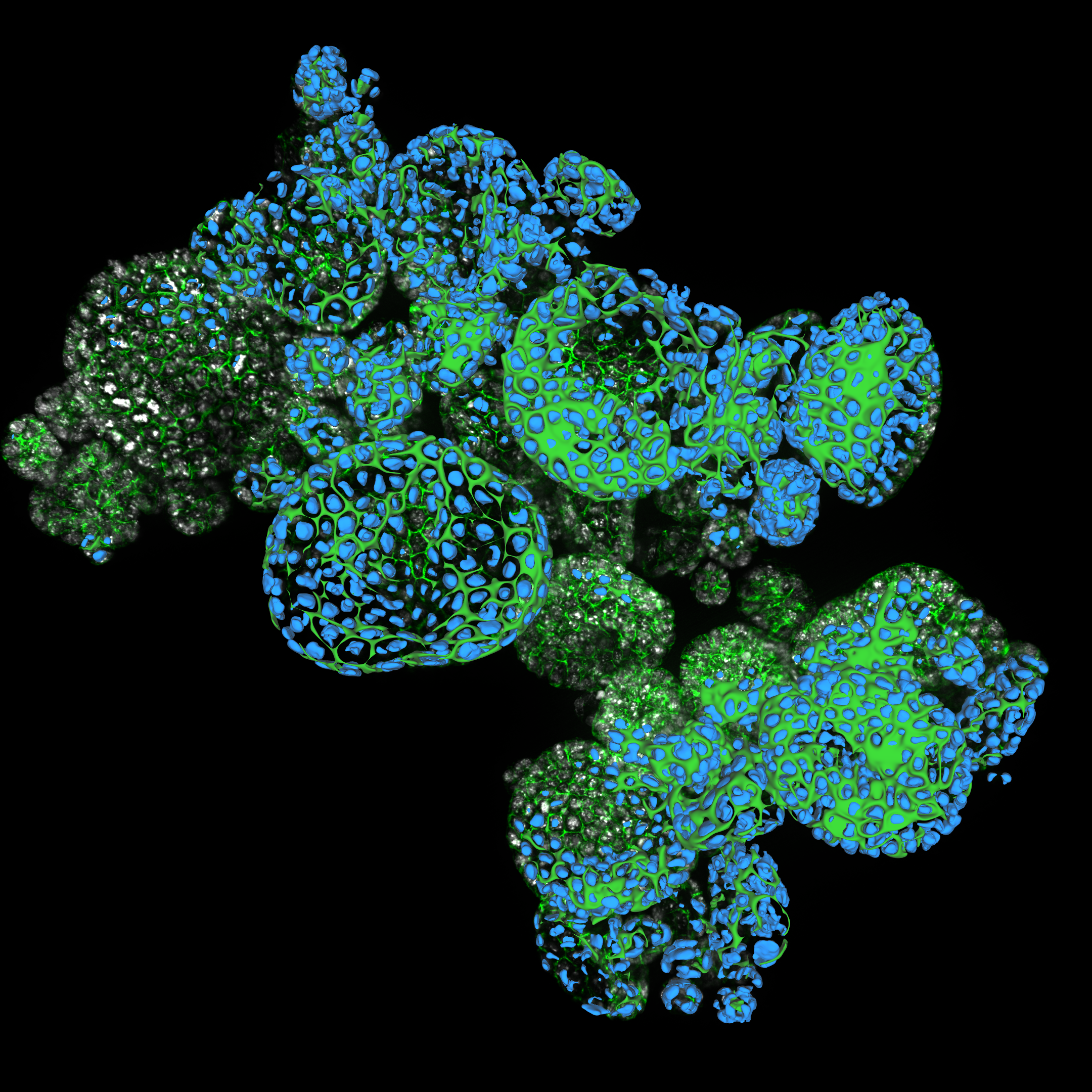
Organoid cluster labelled for nuclei (DAPI, blue) and plasma membrane (GFP, green). Thickness 100 μm. 469 Z planes were acquired using THUNDER Imager Cell (63x objective). Courtesy of M.Sc. Dana Krauß, Medical University of Vienna, Austria.
Organoid models have transformed life science research yet optimizing image analysis workflows remains a challenge. In this webinar, we present a streamlined workflow for organoid imaging, from initial real-time monitoring of 3D cell cultures through to advanced AI-based analysis.
Key webinar learnings:
- How to establish a workflow that combines real-time observations and checks of organoid cultures with detailed 3D imaging followed by accurate segmentation and analysis of the resulting image data
- How to image large sample volumes, such as organoids, at high speed, with low phototoxicity and photobleaching
- How to get cleaner data for accurate AI-based segmentation and quantification of parameters such as growth rate, cell migration, and 3D cellular interactions – enabling deeper insights
Watch the on-demand webinar now
____________________________________________________________________________________________
Imaging Complex Human-Relevant Models for Early Drug Discovery - Technology Showcase
25th June 2026
Join Leica Microsystems and leading experts as they explore how organoids are being used across early drug discovery and translational research workflows, the challenges associated with working with increasingly complex models, and emerging imaging approaches designed to address some of these challenges.